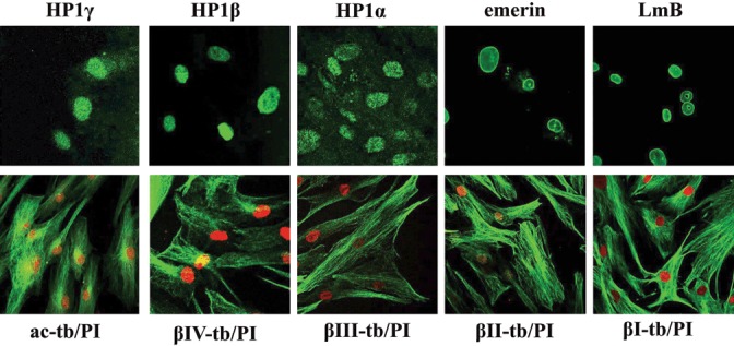
Figure 2.

Expression and localization of tubulin isotypes, proteins of the nuclear envelope and heterochromatin. Immunofluorescence pattern of human mesenchymal stem cells (hMSC) stained with antibodies (green) for lamin B (LmB), emerin, heterochromatin 1 proteins (HP1α, HP1β, HP1γ), β‐tubulin isotypes (βI‐tb, βII‐tb, βIII‐tb, βIV‐tb) and acetylated α‐tubulin (ac‐tb). Nuclei (red) were stained with propidium iodide (PI).
