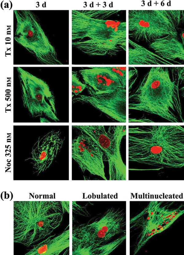
Figure 4.

Effect of taxol and nocodazole on microtubule network and nuclear morphology. (a) Taxol‐ and nocodazole‐treated human mesenchymal stem cells (hMSC) for 3 days (3 d) then grown in drug‐free medium for 3 (3 d + 3 d) and 6 (3 d + 6 d) days, were stained with anti‐tubulin antibodies (microtubules, green) and propidium iodide (nuclei, red). (b) Representative images of multinucleate hMSCs and cells with normal‐looking and with lobulate nuclei (red).
