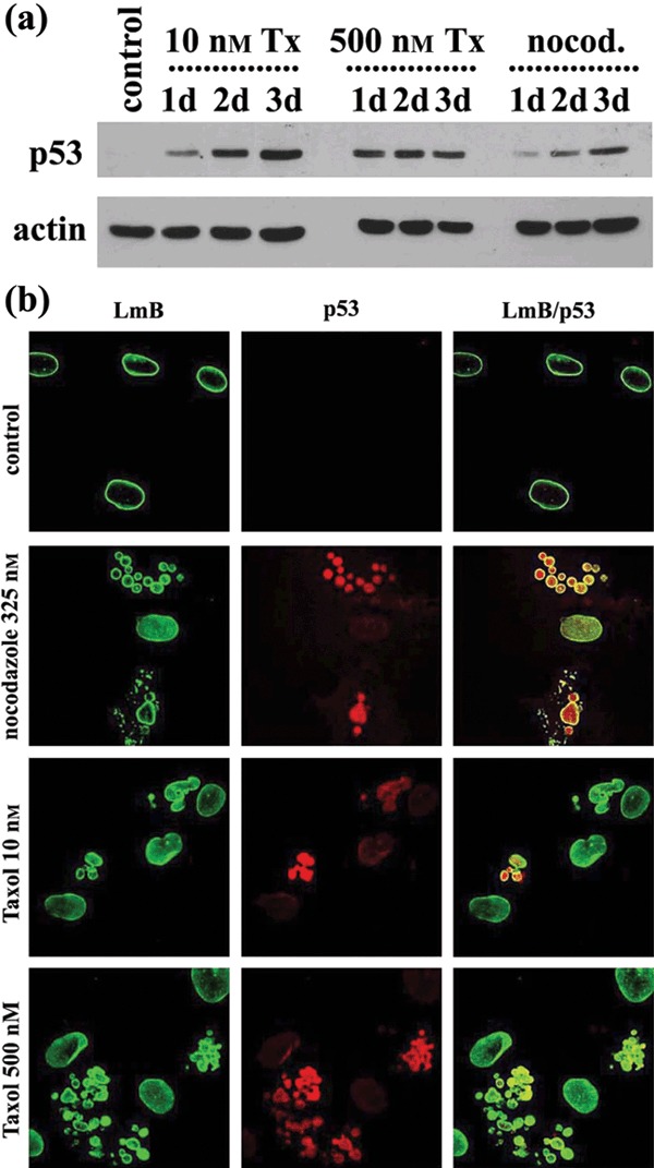
Figure 5.

Expression and localization of p53. (a) Western blot analysis of whole lysates of untreated (control), treated with taxol (Tx) and nocodazole (nocod.) human mesenchymal stem cells (hMSC) for 1 day (1d), 2 days (2d) or 3 days (3d) with p53 and actin antibodies. (b) Typical images of lamin B (LmB) p53 and the merge of two (LmB/p53) in untreated (control) and treated with taxol and nocodazole hMSCs. Note the absence of p53 in untreated cells and its nuclear localization in multinucleate cells, following drug treatment.
