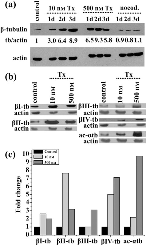
Figure 7.

Effect of taxol and nocodazole on tubulin synthesis. (a) Western blot analysis of whole lysates of untreated (control), treated with taxol (Tx) and nocodazole (nocod.) human mesenchymal stem cells for 1 day (1d), 2 days (2d) or 3 days (3d) with β‐tubulin and actin antibodies. Tubulin/actin (tb/actin) ratios were calculated after determination of intensity of protein bands with image analysis. (b) Western blot of whole lysates of untreated (control) and treated hMSCs with taxol 10 nm or 500 nm, for 3 days with isotype‐specific β‐tubulin, acetylated α‐tubulin (ac‐αtb) and actin antibodies. (c) Histogram depicting changes in tubulin isotype/actin ratios in treated hMSCs with taxol 10 nm or 500 nm for 3 days. Ratios were calculated after determination of intensity of protein bands shown in panel (b) with image analysis.
