
Figure 5.
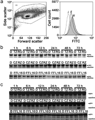
Peroxisome proliferator‐activated receptor (PPAR) agonist‐induced apoptosis is mediated by disruption of mitochondrial membrane potential with concomitant activation of caspase‐9 and caspase‐3. (a) Disruption of mitochondrial membrane potential (ΔΨm) was assessed using the MitoCaptureTM apoptosis detection kit following treatment with ciglitazone (10 µm) in cultured NHU cell populations (left panel) over a period of 6 h (i, vehicle control; ii–iii, 3 and 6 h post‐treatment, respectively). A time‐dependent increase in FITC‐fluorescence (right panel) was evident over 6 h, suggesting disruption of ΔΨm, by decreased ability of the mitochondria to form aggregates of the dye. (b and c) The effects of PPAR agonists troglitazone (TZ; 10 µm), ragagli‐ tazar (RG; 30 µm), ciglitazone (CZ; 10 µm), rosiglitazone (RZ; 10 µm), fenofibrate (FF; 10 µm) and L165041 (L165; 10 µm) on activation of caspase‐9 (b) and caspase‐3 (c) was assessed in cultured NHU cells over a period of 72 h using Western blotting. Full length and active fragments of caspase‐9 were detected at 45 kDa and 38 kDa, respectively. Full length and active fragments of caspase‐3 were detected at 32 and 18 kDa, respectively. All experiments were conducted using at least three independent NHU cultures.