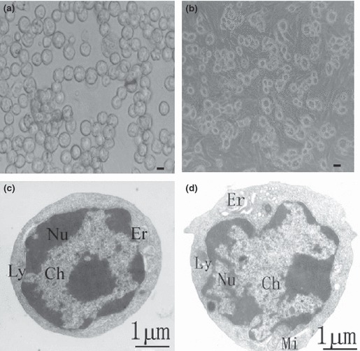
Figure 2.

Morphological features of hepatic oval cells derived from porcine liver. (a, b) Phase‐contrast microscopy showing morphology of the freshly isolated porcine hepatic oval cells (a) and cells cultured for one day (b). Scale bar in a = 3 μm, and bar in b = 6 μm. (c, d) Transmission electronic micrograph showing freshly isolated oval cells from porcine liver with their ovoid nuclei (Nu), condensed chromatin (Ch), high nuclei/cytoplasmic ratio, mitochondria (Mi), endoplasmic reticulum (Er) and lysosome (Ly).
