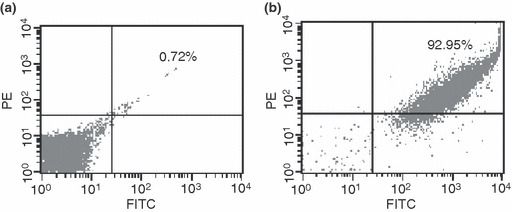
Figure 4.

Double staining and flow cytometric analysis to characterize porcine oval cells as expressing hepatobiliary phenotype. (a) Replacement of primary antibodies with PE‐labelled and FITC‐labelled IgG served as negative control. (b) Freshly isolated cells from porcine liver after incubation with primary antibodies including albumin and CK19, followed by PE‐labelled IgG and FITC‐labelled as secondary antibody.
