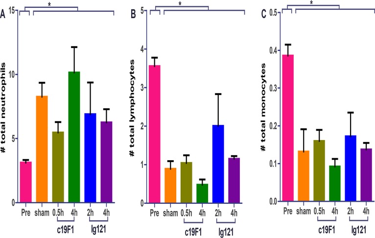
FIG 4.
Cellularity from peripheral blood from all animals (n = 23) prior to SEB challenge (Pre) and 24 h postchallenge and/or treatment with either antibody. Each graph represents neutrophil (A), lymphocyte (B), or monocyte (C) count. All postchallenge values for either sham- or antibody-treated animals were significantly different (*, P < 0.05) from preexposure values. There was no statistical difference between sham control and treatment animals postexposure for any of the values presented.

