Figure 6.

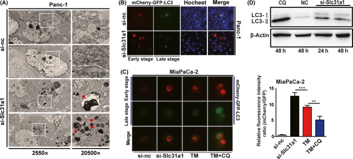
Copper deprivation increased pancreatic cancer cell autophagy. A, the autophagy of Panc‐1 cells that was induced by Slc31a1 interference was presented via transmission electron microscope. B and C, Fluorescence detection of mCherry‐GFP‐LC3 in TM‐treated or Slc31a1 knocked down Panc‐1 and MiaPaCa‐2 cells. Red is representative of an early stage of autophagy, and green quenching indicates the increased function of lysosomes. Data shown are mean ± SD, n = 3, **P < 0.01, ***P < 0.001 (Student's t test). D, Western blot detected LC3I and LC3II in si‐Slc31a1‐transfected Panc‐1 cells
