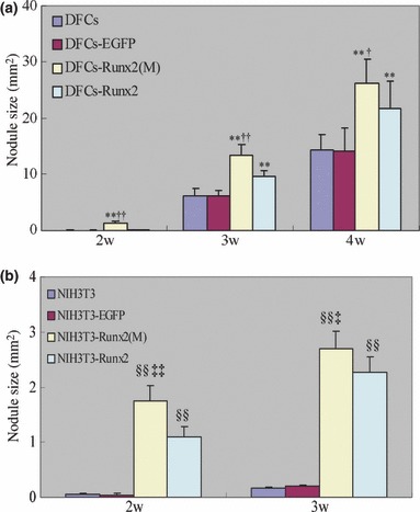
Figure 5.

Effects of Runx2 overexpression on mineral nodule formation. Cells were cultured in mineralizing media and von Kossa staining was performed to evaluate the mineral nodule formation on week 2, 3, 4. The area covered by mineral nodules in each well was determined. The data were presented as the mean ± SE for three experiments. **P < 0.01 vs. DFCs and DFCs‐EGFP, †P < 0.05 vs. DFCs‐Runx2, ††P < 0.01 vs. DFCs‐Runx2, §§P < 0.01 vs. NIH3T3 and NIH3T3‐EGFP, ‡‡P < 0.01 vs. NIH3T3‐Runx2, ‡P < 0.05 vs. NIH3T3‐Runx2. Statistical significance was determined using t‐test.
