Figure 1.

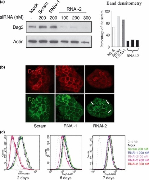
Dsg3 knockdown in HaCaTs. (a) Western blotting of Dsg3 in HaCaT cells transfected either with or without scrambled (Scram), RNAi‐1 and RNAi‐2 siRNAs at different concentrations, for 2 days; loading control –β‐actin. Densitometry of Dsg3 blot is shown on the right. Note that Dsg3 levels in RNAi‐2 treated cells were ∼20% of that of scrambled control regardless of different siRNA concentrations. (b) Confocal microscope images of HaCaT cells dual labelled for Dsg3 (AHP319) and Dp (115F) following transfection of scrambled siRNA, RNAi‐1 and RNAi‐2 for 3 days. Disruption of desmosomal junctions was seen in RNAi‐2‐treated cells for Dp staining (arrows). (c) FACS analysis of Dsg3 expression, labelled with mouse Dsg3 Ab 5H10, in HaCaTs transfected similarly (a) for a period of up to 7 days. Again, significant knockdown of Dsg3 was seen after 2 days regardless of siRNA concentration for RNAi‐2. RNAi‐1 showed very subtle effect on Dsg3 expression (blue line). After 7 days, expression of Dsg3 in RNAi treated cells recovered to almost that of normal control levels.
