
Figure 5.
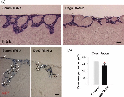
Cells with Dsg3 silencing displayed poor quality of skin regeneration. (a) One week organotypic culture of cells with or without Dsg3 knockdown (n > 3). Top: H&E staining; Bottom: Ki67 staining, positive cells marked with white arrows. Bars, 50 μm. (b) Quantification of epithelial areas in organotypic culture sections (n = 12). Note that cells treated with RNAi produced significantly lower cellular mass (*P < 0.05) and less Ki67 positive cells.