Figure 6.

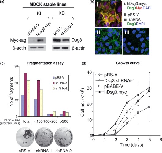
Modulation of Dsg3 expression affected cell proliferation in MDCK cells. (a) Western blotting of cell lysates extracted from cells with either overexpression (knock‐in: KI) or knockdown (KD) of Dsg3. (b) Confocal microscopy of MDCK cells with ectopic Dsg3 expression [myc‐tag in (i)] or with Dsg3 knockdown (iii). Images in (ii) were vector control cells. (c) Dispase fragmentation assay of MDCK cells with either vector or Dsg3 shRNAi transduction. Cells were grown to confluence before being treated with 2.4 units/ml dispase for about 30 min to detach epithelial sheets. These sheets were subjected to mechanical stress by pipetting five times with 1 ml tips. Epithelial fragments were quantified by ImageJ and increased fragments (up to 2‐fold) were seen in cells with Dsg3 silencing by shRNAi‐1 or shRNAi‐2, respectively. Data are averages of duplicates in each group. (d) Growth curve of matched MDCK cells with up‐ or down‐regulation of Dsg3. Cells with overexpression had higher proliferation, but those with Dsg3 knockdown exhibited lower growth rate compared to matched control cells.
