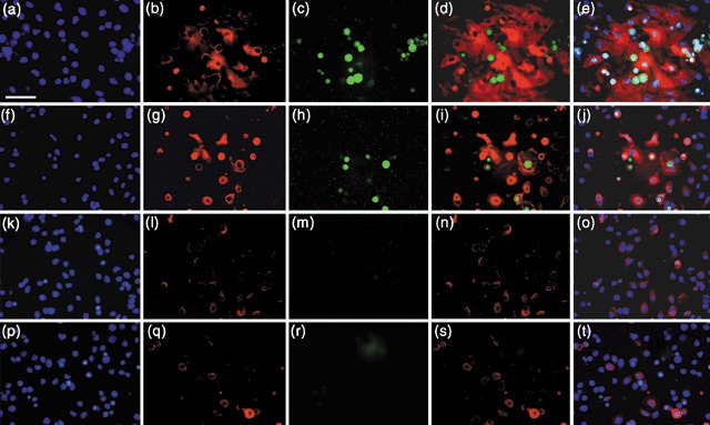
Figure 5.

Expression of BMPR1A and Oct‐4 in keratinocytes following exposure to CM or recombinant BMP4. (a–e) Keratinocytes cultured in 1 : 1 DKSFM : CM for 24 h. (a) DAPI (4′,6‐diamidino‐2‐phenylindole); (b) anti‐BMPR1A; (c) anti‐Oct‐4; (d) merge BMPR1A and Oct‐4; (e) merge BMPR1A, Oct‐4 and DAPI. (f–j) Keratinocytes cultured in 1 : 1 DKSFM : uCM +100 ng/mL rBMP4, for 24 h. (f) DAPI; (g) anti‐BMPR1A; (h) anti‐Oct‐4; (i) merge BMPR1A and Oct‐4; (j) merged BMPR1A, Oct‐4 md DAPI. In a–j, note co‐staining of membrane BMPR1A and Oct‐4, and ESC‐like rounded morphology. Additionally, note small, rounded cells with bright cytoplasmic BMPR1A, but no Oct‐4, suggesting BMPR1A is expressed before Oct‐4. (k–o) Control 24 h DKSFM cultures. (k) DAPI; (l) anti‐BMPR1A; (m) anti‐Oct‐4; (n) merge BMPR1A and Oct‐4; (o) merge BMPR1A, Oct‐4 and DAPI. (p–t) Control 24 h 1 : 1 DKSFM : uCM cultures. (p) DAPI; (q) anti‐BMPR1A; (r) anti‐Oct‐4; (s) merge BMPR1A and Oct‐4; (t) merge BMPR1A, Oct‐4 and DAPI. Note, no Oct‐4 or membrane BMPR1A staining in controls. All images, ×40. Scale bar = 50 µm.
