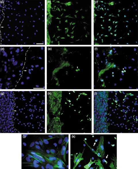
Figure 8.

Expression of nestin, NeuN and K14 in cells grown for 7 days in neuronal differentiation medium. Keratinocytes were exposed to CM for 24 h then cultured in neuronal differentiation medium for 7 days. Dashed line marks border between keratinocyte colony and emerging neuronal‐type cells. Arrows indicate direction of neuron‐like cell migration. (a–c) Cultures stained with antibody to nestin. ×20 magnification. (a) DAPI (4′,6‐diamidino‐2‐phenylindole); (b) anti‐nestin; (c) merged. (d–f) Cultures stained with antibody to NeuN. ×40 magnification. (d) DAPI; (e) anti‐NeuN; (f) merged. Arrowheads mark NeuN+ nuclei. Note, multilayered colony edges showed non‐specific accumulation of NeuN antibody. (g–i) Cultures stained with antibody to keratin 14 (K14). ×20 magnification. (g) DAPI; (h) anti‐K14; (i) merged. Note, in the few K14+ neuronal‐type cells, the K14 filaments appeared collapsed around the nuclei. (j) ×60 magnification of antinestin/DAPI. (k) ×60 magnification of anti‐K14/DAPI. Arrows point to perinuclear clumps of keratin in cells. Scale bars = 50 µm.
