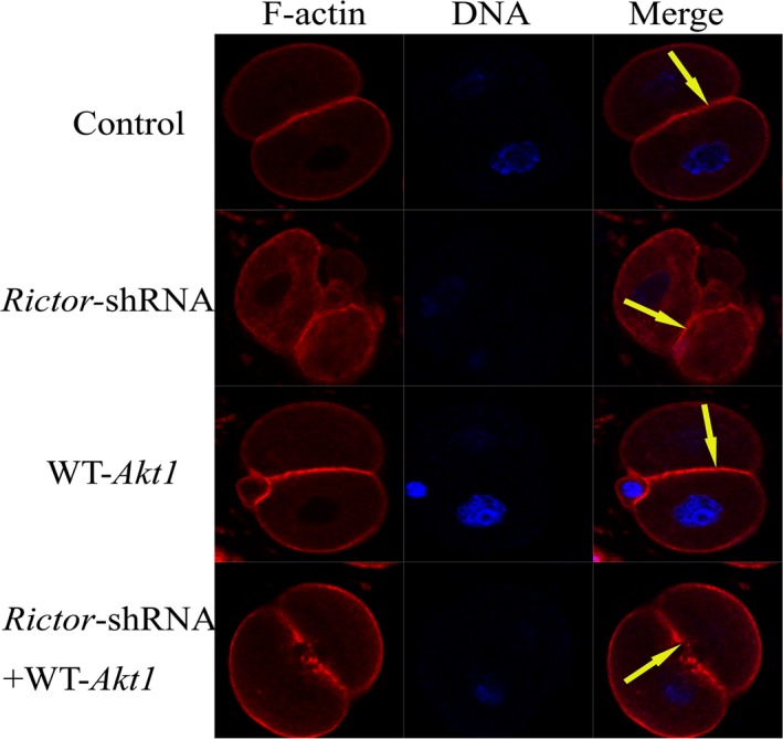
Figure 3.

The mTORC2/Akt1 pathway rearranges the F‐actin cytoskeleton of one‐cell stage fertilized eggs. Microinjection of Rictor shRNA then with myr‐Akt1 mRNA into mouse one‐cell stage embryos. Staining for F‐actin (red) revealed the organization of the F‐actin cytoskeleton in mouse fertilized eggs (F‐actin is shown by the yellow arrow). Scale bar: 20 μm
