Figure 4.

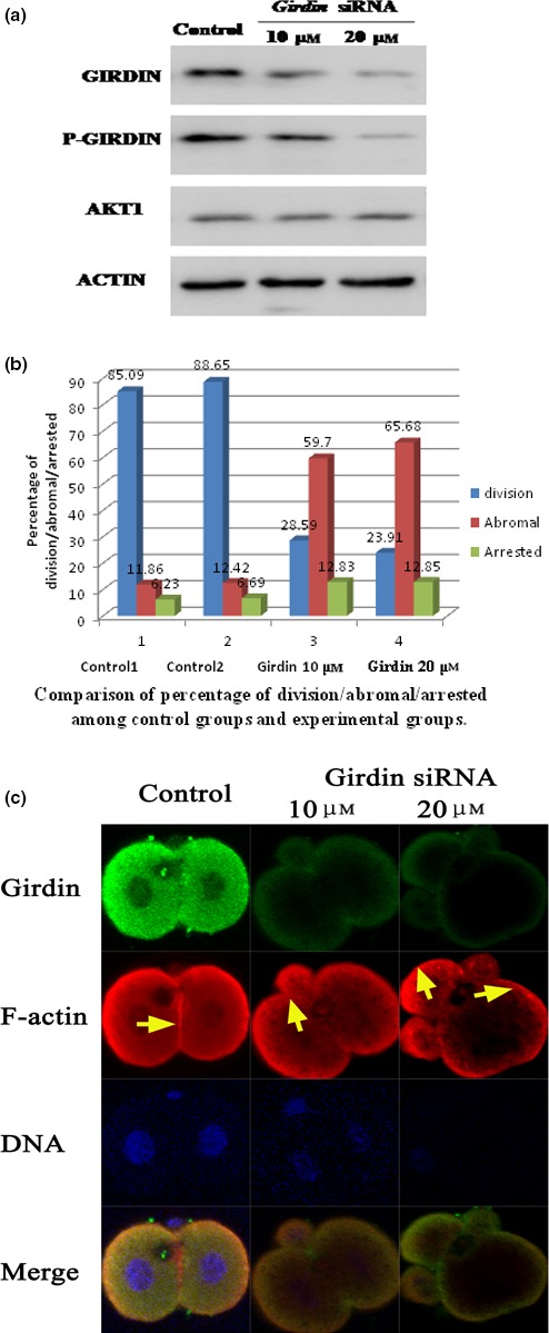
Girdin is essential for rearrangement of the F‐actin cytoskeleton and the development of mouse fertilized eggs. (a) Depletion of Girdin in mouse one‐cell staged fertilized eggs by siRNA. Total cell extracts from control siRNA‐ and Girdin siRNA‐injected one‐cell staged fertilized eggs were subjected to Western blot analyses and immunodetection with anti‐Girdin, anti‐p‐Girdin, anti‐Akt1 and anti‐actin antibodies. (b) The cleavage rate in cultured mouse embryos after Girdin siRNA injections shows that the total number of eggs undergoing cell division is given above each bar graph from three independent experiments. (c) F‐actin cytoskeleton of embryos derived from fertilized eggs treated with Girdin siRNA. Mouse fertilized eggs were treated with control or Girdin siRNAs and fixed 48 hours later, followed by staining with rhodamine‐phalloidin and anti‐Girdin antibody. In situ validation of the interaction between Girdin and polymerized F‐actin by confocal microscopy is shown. One‐cell stage mouse fertilized eggs were treated with 10 or 20 μmol/L Girdin siRNA. Immunolocalization of Girdin is revealed by green staining (antibody), and immunolocalization of actin is revealed by red staining. Control fertilized eggs (21 hours after hCG): one‐cell arrested embryos derived from fertilized eggs that were injected with Girdin siRNA 21 hours after hCG and cultured for 24 hours; control two‐cell embryo: in control fertilized eggs, F‐actin forms a regular ring in the cell cortex. In embryos derived from fertilized eggs treated with girdin siRNA, polymerized F‐actin was observed in the cytoplasm and formed irregular patches that were scattered randomly in the cortex. Scale bar: 20 μm
