Figure 5.

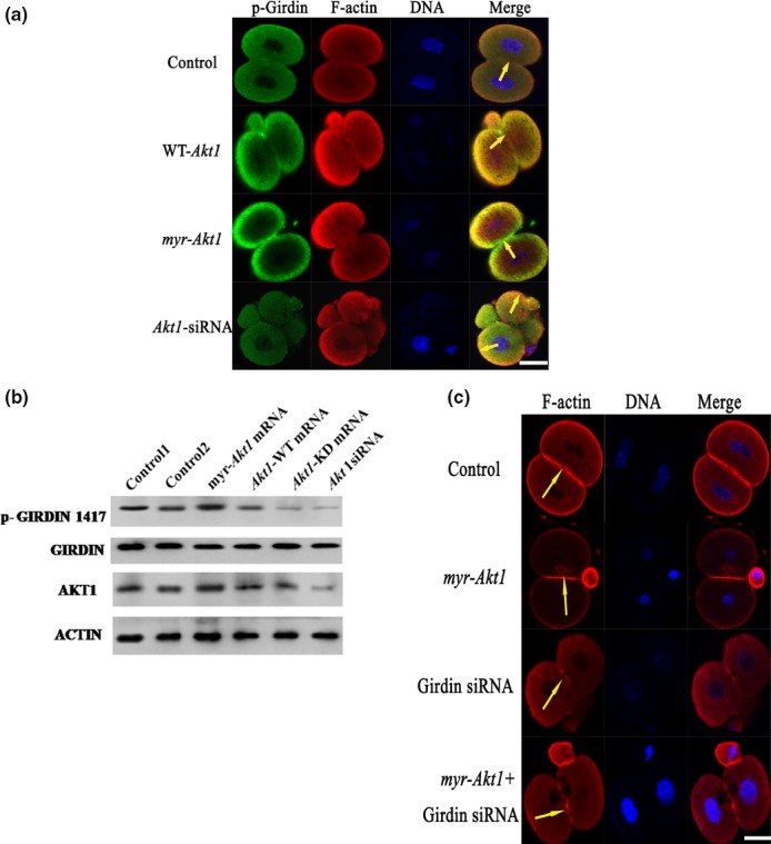
The Akt1/ Girdin pathway regulates rearrangement of the F‐actin cytoskeleton of one‐cell stage fertilized eggs. (a) One‐cell stage mouse embryos were first microinjected with Akt1‐WT mRNA, myr‐Akt1 mRNA or siRNA, then stained with rhodamine‐phalloidin (20 μmol/L; actin labelling as shown in red) and anti‐P‐Girdin (1:50; Girdin labelling; as shown in green). Merged colour detection between P‐Girdin and polymerize F‐actin appears as yellow stained images. The areas of co‐localization are highlighted with yellow arrowheads. Scale bar: 20 μm. (b) 200 fertilized eggs were treated with mRNA coding for Akt1‐WT, myr‐Akt1, Akt1‐KD or siRNA against Akt1. Western immunoblot analyses assessed following immunoreaction with anti‐P‐Girdin (upper panel) and anti‐Girdin (lower panel) antibodies are shown. (c) One‐cell stage mouse embryos were first microinjected with 0.03 ng of myr‐Akt1 mRNA as described in the Materials and Methods, and then 1–2 hours later with Girdin siRNA, following which specimens were stained with rhodamine‐phalloidin (20 μmol/L; actin labelling shown in red). Scale bar: 20 μm
