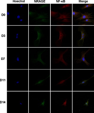
Figure 7.

Immunofluorescence staining of NRAGE and NF‐κB relocation in mDPCs during odontoblastic induction for days 0, 3, 7, 11 and 14 (400× magnification). Immunofluorescence staining with anti‐NRAGE antibody (green), anti‐NF‐κB antibody (red) and Hoechst (blue) was performed. Negative control staining was performed with immunoglobulin G control antibody.
