
Figure 3.
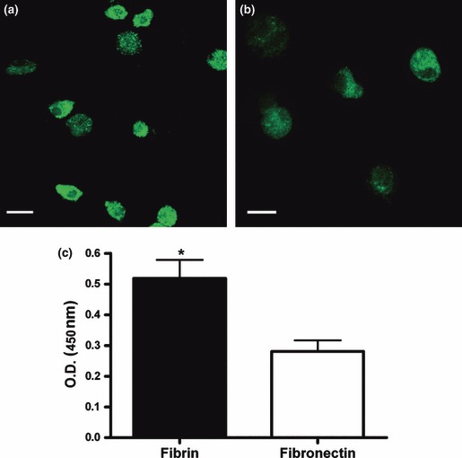
Cell viability. Cells cultured for 1 week on fibrin (a) or fibronectin (b) were incubated with calcein‐AM. Viable cells are evidenced by the presence of green staining in cell cytoplasm. Representative confocal laser scanning microphotographs are shown (n = 3). Scale bar = 10 μm. (c) Cells cultured for 1 week on fibrin or fibronectin were incubated with the tetrazolium salt WST‐1 for 4 h at 37 °C to produce a formazan dye, quantified by measuring the optical density at 450/655 nm. *P < 0.05. Mean ± SEM of three different experiments.