Figure 3.

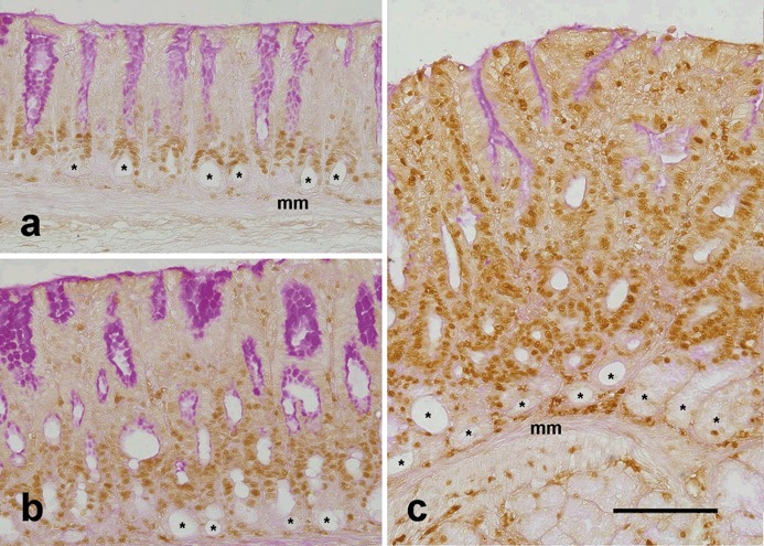
Labelling of dividing cells in control (a) and TFF1‐deficient (b, c) mice at ages of 1 month (b) and 2 months (a, c). Tissue sections were probed with polyclonal anti‐proliferating cell nuclear antigen (PCNA) antibody and then were incubated with biotinylated anti‐rabbit immunoglobulin G. Following incubation with peroxidase‐conjugated avidin, antigen‐antibody binding sites were visualized by adding diaminobenzidine colouring agent. Nuclei of proliferating cells in the G1 or S phase of the cell cycle appear brown because they express a high level of PCNA. Note that in control tissue (a), proliferating cells occur in isthmus regions at the pit‐gland borders. Also note the round lumina (asterisks) of the glands at the bottom of the mucosa, next to the muscularis mucosa (mm). These lumina are lined with terminally differentiated post‐mitotic gland mucous cells which are not labelled with PCNA. In TFF1 knockout tissues (b, c), the mucosa is thick and PCNA‐labelled proliferating cells are numerous, as compared to those of control tissue in (a). The three tissue sections were counterstained with periodic acid Schiff (PAS). PAS‐positive cells are seen at the luminal surface and along the pit walls, especially in (a) and (b). Note that PCNA‐labelled cells in control and knockout tissues show little or no PAS‐positive staining. Bar = 90 µm (a–c).
