
Figure 4.
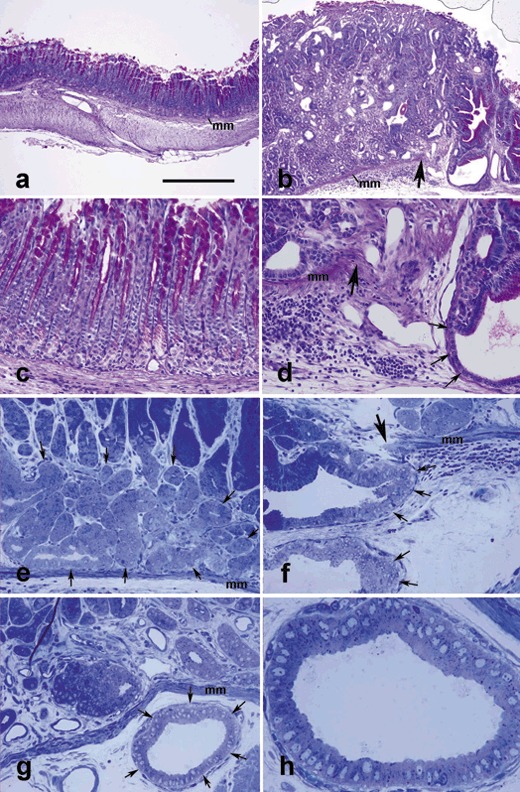
Antral mucosal tissue sections of control (a, c) and TFF1 knockout (b, d–h) mice at 12 (a–d) and 17 (e–h) months of age, stained with periodic acid Schiff (PAS) and haematoxylin (a–d) or toluidine blue (e–h). In control tissue (a), the muscularis mucosa (mm) separates the epithelial units above from submucosa and muscularis externa below. In (b), note that the epithelial units are simple and tubular and apices of cells lining pit regions are PAS‐positive (purple). In TFF1 knockout tissue, note increased thickness of the mucosa (b versus a) and decreased amount of PAS‐stained mucus, as compared to control tissue in (a). Also note invasive epithelial profiles (b, d, f–h), penetrating through the muscularis mucosa (mm) which is lacking at the right (in b, d) or left (in f) of the large arrow. In 0.5‐µm‐thick plastic sections of the basal portion of the pyloric antral mucosal tissues, amplified progenitor cells form a large group of pale cells (surrounded by arrows in e) which appear similar to a carcinoma in situ, or they cross the muscularis mucosa and invade the submucosa to form multiple cyst‐like profiles (arrows in f and g). High magnification of the invasive profile of (g) is shown in (h). It illustrates the small size of the invasive cells with little or no visible secretory dense granules. Bar = 300 (a, b), 90 (c–g), and 35 (h) µm.