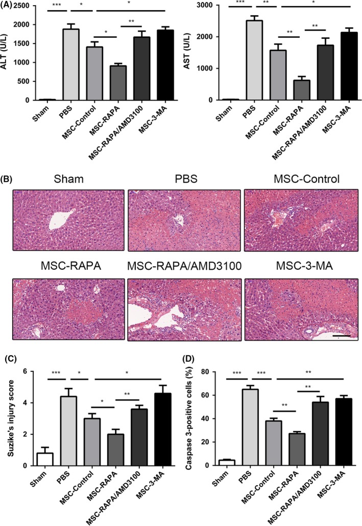
Figure 4.

Enhancement of autophagy in UC‐MSCs is shown to protect the liver by decreasing levels of serum biomarkers and the histological features of hepatic injury after ischaemia/reperfusion (I/R) injury in vivo. A, Serum alanine and aspartate aminotransferase levels were detected after I/R injury in each treatment group. Data are shown as the mean ± standard error of the mean (n = 6 mice/group). B, Haematoxylin and eosin staining of liver tissues in each group to assess the amount of liver damage after I/R injury. Scale bar: 200 µm. C, Suzuki's injury score for each group calculated by randomly selecting five fields in each tissue sample. Results of statistical analysis are presented as the mean ± standard error of the mean (n = 6 mice/group). D, Amount of caspase‐3 in each group was assessed using immunohistochemistry to determine the percentage of caspase‐3‐positive cells in the liver. Statistical analysis was performed to determine the number of caspase‐3‐positive cells. Data are shown as the mean ± standard error of the mean (n = 6 mice/group). *P < 0.05, **P < 0.01, ***P < 0.001 (one‐way analysis of variance). UC‐MSCs, umbilical cord‐derived mesenchymal stem cells
