
Figure 5.
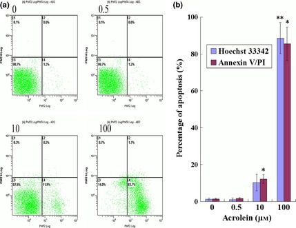
Acrolein ( Acr )‐induced apoptosis. Cells were exposed to various concentrations of Acr for 12 h, stained with an FITC‐labelled annexin V/PI apoptosis kit and measured by flow cytometry (a and b), stained with Hoechst 33342 measured by fluorescence microscopy (b). There were increased numbers of apoptotic cells compared to lower concentration Acr. Apoptosis was the reason for cell viability decrease treated by Acr. *P < 0.05, compared to 0 μm and stained with FITC‐labelled annexin V/PI. **P < 0.05, compared to 0 μm and stained with Hoechst 33342.