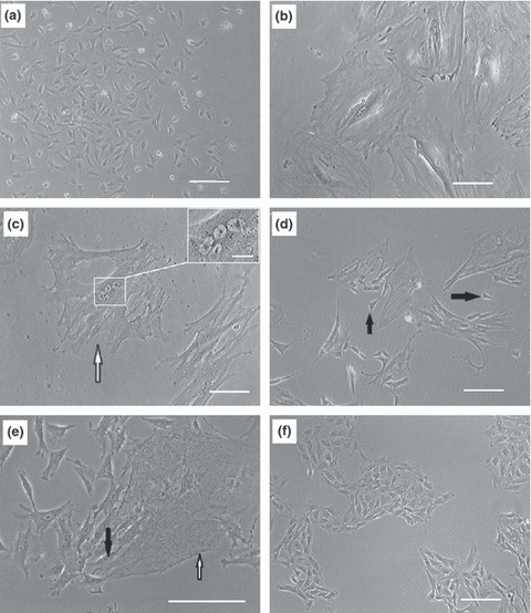
Figure 1.

Morphology of rabbit BM‐MSCs in different phases of population growth. Bone marrow contents were transferred to flasks and after 24 h, non‐adherent cells were discarded. Remaining cells were expanded and after reaching 50% confluence, they were detached and replated as passage 1 cells (a). Proliferation rate of the cells gradually descended until they stopped dividing after 3.2 ± 1.3 passages. At this point, cells became large (b) and subsequently, several nuclei were visible in each large cell (arrow head), nuclei at higher magnification (inset scale bar: 20 μm) (c). After 35 ± 7 days in the dormant phase, each large multinucleate cell broke into several small cells (d and e). White arrow shows a large multinucleate cell and black arrows demonstrate newly formed small cells. Small cells proliferated constantly and rapidly over extended periods (f). Scale bars: 100 μm.
