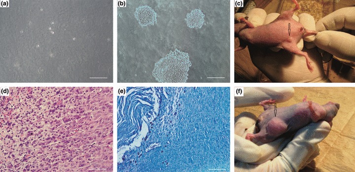
Figure 6.

Assessment of in vitro and in vivo tumour formation potential of rabbit BM‐MSCs. MSC could not form colonies in soft agar colony formation assay (a), whereas under the same conditions, MKN45 cells (used as positive controls), formed colonies (b). In mice transplanted with immortal MSCs, within a few days, small nodules became visible at the transplantation site (c). This nodule disappeared by day 8. Histopathological examination of H&E‐stained slides (d) showed that cells were plump to spindle‐shaped with merging eosinophilic cytoplasm. In the centre, cytoplasm of cells showed hydropic clear changes and at the left, necrosis was seen. Apoptotic cells were also noted. Mast cells were arranged at the periphery of the mass in Giemsa‐stained samples (e). No indication of neoplastic change was seen. MKN45 cells formed tumours at the transplantation site (arrowhead) (f). Scale Bars: 100 μm.
