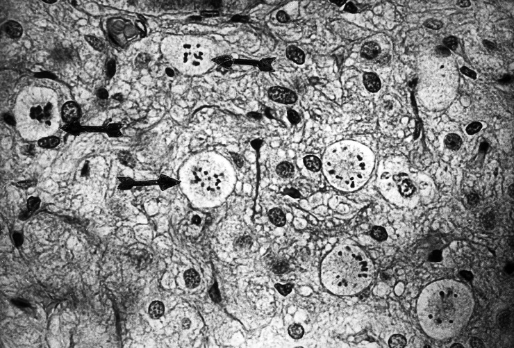
Figure 1.

A small portion of the neural lobe with eight mitotic figures, from a nephrectomized rat treated with a single dose of LiCl. Arrows on the left side point to three mitoses. The right side has five cells in mitosis. Individual chromosomes are clearly visible. This unusual clustering of mitoses supports the suggestion that some pituicytes may divide more than once after stimulation. This single 5 µm section had 31 mitoses in all, in an area of 1.23 mm2. In contrast, neural lobes from control rats usually had no mitoses in the entire section. Hematoxylin and eosin (magnification × 400).
