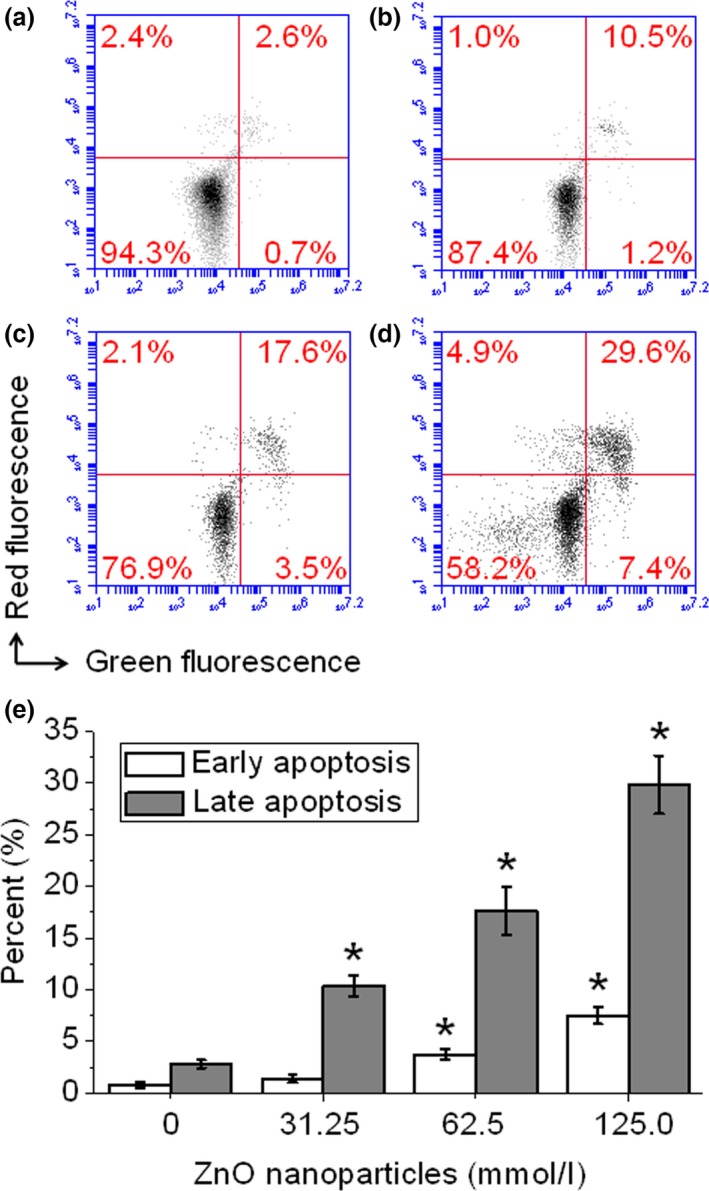
Figure 5.

ZnO nanoparticle‐induced cell apoptosis and/or necrosis monitored by flow cytometry. Cells were treated with 0, 31.25, 62.5 and 125.0 μmol/l of ZnO nanoparticles and cultured for 24 h, then all cells were collected, washed with cold PBS and stained with annexin V/PI double staining solution, finally all samples were determined by flow cytometry. (a) Untreated cells; (b) cells treated with 31.25 μmol/l of ZnO NPs; (c) cells treated with 62. 5 μmol/l of ZnO NPs; and (d) cells treated with 125.0 μmol/l of ZnO NPs. Measurements were repeated three times and NPs = nanoparticles. *P < 0.05 versus relevant control samples.
