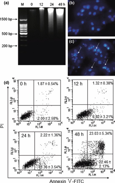
Figure 1.

AML induced apoptosis in K562 cells. DNA fragmentation was checked by ethidium bromide staining (a). Cells were cultured with 20 μg/ml AML for 0, 12, 24 and 48 h respectively, ‘M’ represents marker. Morphological observation of nuclei was visualized using a fluorescence microscope and DAPI staining (400×). Cells were incubated in the absence (b) or presence (c) of 20 μg/ml AML for 24 h. Arrows indicate apoptotic features (condensed chromatin and nuclear fragmentation). Apoptosis of K562 cells was quantified using annexin V/PI staining and flow cytometry (d). Cells were treated with 10 μg/ml AML for 0, 12, 24 and 48 h respectively. Data expressed as mean ± SD, obtained from experiments performed in triplicate.
