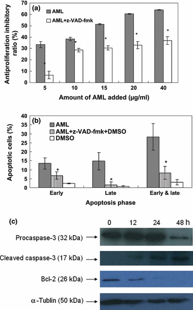
Figure 2.

AML induced apoptosis through a mitochondria‐mediated and caspase‐dependent pathway. Cells were incubated in a variety of concentrations of AML and 40 mm z‐VAD‐fmk, for 48 h: cell viability was checked by MTT assay (a). Cells were incubated in 10 μg/ml AML and 40 mm z‐VAD‐fmk for 48 h; apoptosis was investigated using flow cytometry (b). Data expressed as mean ± SD, obtained from experiments performed in triplicate, *P < 0.05 versus AML group. The K562 cells were treated with 10 μg/ml AML for the variety of time periods, and levels of Bcl‐2 and caspase‐3 were detected by western blot analysis (c). α‐tublin was used as equal control, and results are representative of experiments performed in triplicate.
