
Figure 3.
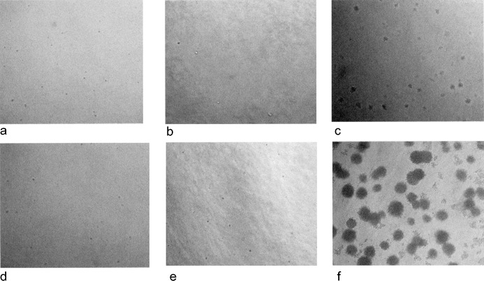
Culture of mesenchymal stromal cells (MSCs) on soft agar for the assay of anchorage dependence. Pictures were taken on day 7 (a, b, c) and 14 (d, e, f) and correspond to ΗL‐60 (c, f), P2 MSCs (a, d) and P6 MSCs (b, e). No colonies were observed in MSC samples in contrast with HL‐60 cells where foci were visible from day 7.