
Figure 8.
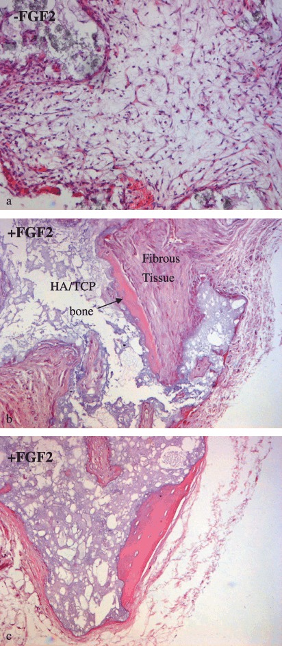
Histological analysis. Deposition of bone matrix onto the carrier in transplants of mesenchymal stromal cell (MSC) grown with fibroblast growth factor (FGF)‐2 (b, c) is shown. Only fibrous tissue and hydroxyapatite/tricalcium phosphate (HA/TCP) particles were detected in transplants of untreated cells (a). None of the samples showed evidence of tumour growth.