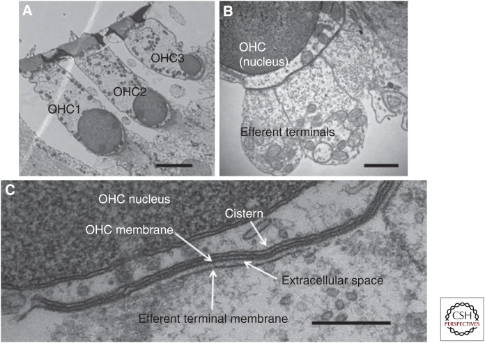
Figure 3.
Synaptic ultrastructure in outer hair cells (OHCs) of mice. (A) Transverse section showing an OHC of each row. (B) Higher magnification showing three efferent terminals on one OHC. (C) Postsynaptic cistern of OHC, aligned with efferent terminal. Scale bars, 4 µm (A); 1 µm (B); 250 nm (C). (From Fuchs et al. 2014; adapted, courtesy of HHS Public Access.)

