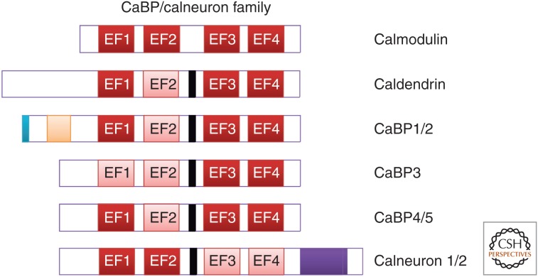
Figure 8.
Schematic diagram showing the domain structure of calmodulin and members of the CaBP/calneuron protein family. Active EF-hand motifs are shown in red and inactive EF-hand motifs are shown in pink. Compared to calmodulin the calcium-binding proteins (CaBPs) have an extended linker region between their first EF-hand pair and their second EF-hand pair (shown in black). CaBP1 and CaBP2 have an N-myristoylation site (shown in blue). CaBP1 and CaBP2 have alternative splice sites at their amino terminus, which give rise to long and short isoforms (shown in orange). Calneurons 1 and 2 possess a 38 amino acid extension at their carboxyl terminus (shown in purple).

