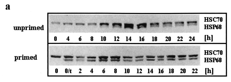
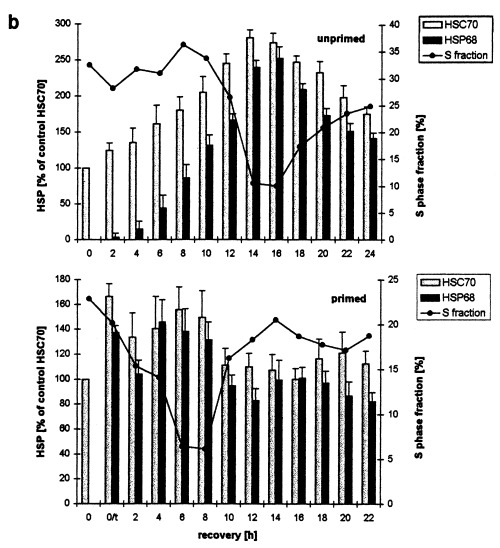
Figure 4.


Western blot determination of HSC70 and HSP68 levels in asynchronously proliferating C6 cells after heat shock. Primed and unprimed cells (0 h) were heat shocked (30 min, 44 °C), harvested after the indicated recovery times at 37 °C and analysed by western blot. (a) Representative immunodetection using the SPA‐820 antibody that recognizes HSC70 as well as HSP68. (b) Western blots of unprimed (top panel) and primed C6 (lower panel) were quantitatively evaluated by video scanning measurement using the CREAM research software (Ver 4.1). Values of HSC70 (grey columns) and HSP68 (black columns) are relative amounts of pixel density given in percentage of the HSC70 value of unstressed, unprimed C6 cells (left ordinate) and are plotted against the recovery time. The fraction of cells in S phase is displayed as a line (right ordinate, see Figure 1 for other fractions). 0/t: primed C6 cells directly before the second heat shock. Values are means (±SE) of at least three independent experiments.
