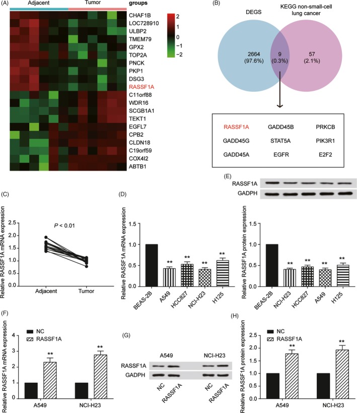
Figure 1.

Characterisation of RASSF1A expression and construction of the abnormally expressed non‐small‐cell lung cancer (NSCLC) cell lines. A, Heat map: Among 20 genes, RASSF1A was significantly lowly expressed in cervical cancer tissues compared with that of adjacent tissues. B, Venn diagram of two sets: DEGS and KEGG non‐small‐cell lung cancer. RASSF1A was chosen from nine intersections. C, qRT‐PCR results showed that RASSF1A expression in tumours was significantly lower than that in adjacent tissues. D, RASSF1A expression in the NSCLC and normal cell lines by qRT‐PCR. E, Western blot results for RASSF1A expression in NSCLC and normal cell lines. F, RASSF1A was overexpressed in A549 and NCI‐H23 cell lines by RT‐qPCR. G, RASSF1A was overexpressed in A549 and NCI‐H23 cell lines by Western blot. H, The Western blot results in a bar graph. The data are from one representative experiment among three that were performed identically and are expressed as the means ± SD. **P < 0.01
