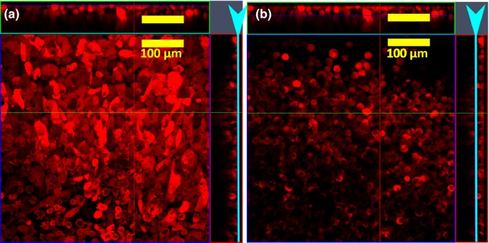
Figure 4.

Z‐stack analysis of a 3D culture of PC 3‐mOrange cells assembled by magnetic forces. (a) Cells on the lower end of the cluster that were in contact with the glass substrate of the coverslip show the familiar elongated form of monolayer culture, while (b) cells which have physical contact only with other cells acquired a spherical morphology. The light blue arrowhead and the line indicates the position in the Z‐plane for the X‐Y image shown.
