Figure 6.

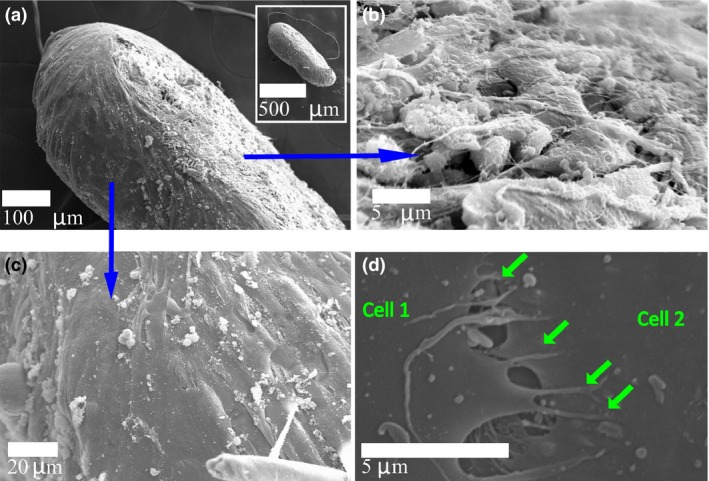
Human lung fibroblast ( HFL ‐1) cells self‐organized into a tight spheroid. (a) Side view of a 3D cluster (inset: lower magnification) shows the flattened‐spheroid shape assumed by the HFL‐1 cells. (b) The basolateral surface of the cluster that was in contact with the glass coverslip contained cells with a rounded morphology. Several inter‐cellular contacts aided by extra‐cellular fibers are seen. (c) The apical surface, which formed the interface between the cluster and the culture medium, was smooth and comprised of skin‐like stretched cells. (d) The stretched cells formed inter‐cellular contacts through filopodia‐like protrusions (arrows).
