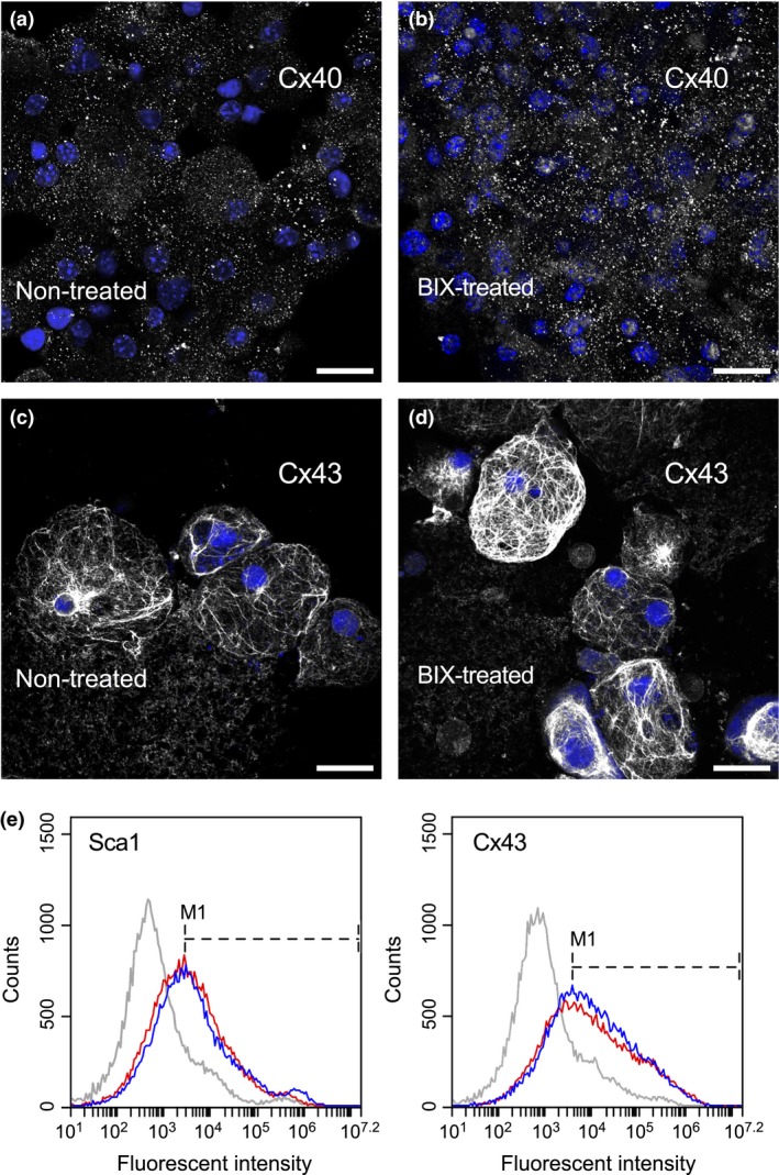
Figure 7.

Cell membrane marker expression by phase bright cells. PBCs collected from either (a,c) non‐treated control or (b,d) BIX01294‐treated atrial cultures were immunostained (grey) for (a,b) Cx40 or (c,d) Cx43, and counterstained with DAPI (blue). Neither gap junction protein showed any comparative dissimilarities on their pattern of expression among the two culture conditions. Scale bar = 20 μm. (e) Flow cytometric analysis of Sca1 and Cx43 showed comparable histogram profiles. Plots for non‐treated control and BIX01294‐treated PBCs are shown in red and blue, respectively, with the grey lines representing cells stained with secondary antibody only. Placement of the M1 marker at peak fluorescence for each antigen indicate that Sca1‐positive cells represent 51.1% and 53.2%, and Cx43‐positive cells represent 61.0% and 65.0% of non‐treated and BIX01294‐treated populations of phase bright cells, respectively.
