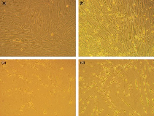
Figure 2.

Morphology of ADSCs, dADSCs and SCs by phase‐contrast microscopy. (a) ADSCs cultured alone showed flattened fibroblast‐like morphology; (b) ADSCs co‐cultured with Schwann cells for 6 days differentiated to a Schwann cell phenotype; (c) Morphology of treated cells was exactly the same as SCs, when co‐cultured for 21 days; (d) Images of purified Schwann cells (magnification: ×100).
