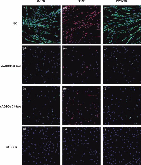
Figure 5.

Immunofluorescence staining in Schwann cells, dADSCs and uADSCs. (a, d, g, j) S‐100; (b, e, h, k) GFAP and (c, f, i, l) P75NTR. Nuclei labelled blue with DAPI. Morphology of dADSCs: purity of isolated Schwann cells was more than 99% (magnification: ×100).
