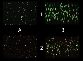
Figure 5.

Representative lateral and vertical views of confocal image stacks obtained from live/dead‐stained gel slices. Both images are taken from the bioconstruct interior, at day 0 in the upper row, and at day 7 in the lower. The images in column A are vertical views, in column B lateral views. Lateral views are software rendered from about 100 individual vertical‐view images. Green objects are stained by calcein AM, red by ethidium homodimer. Yellow objects represent structures that are stained by both ethidium and calcein. The main contribution to the observed reduction in viability over time came from an increased fraction of cells stained with both markers.
