
Figure 3.
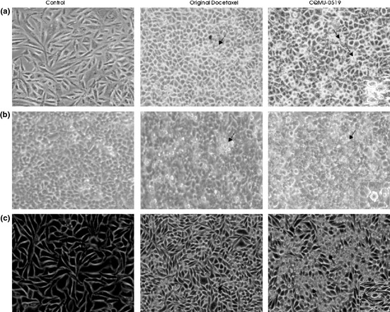
Light microscopy with 100% white balance. (a) A549, (b) SKVO3 and (c) MCF7 cells were treated with our reagents, similar incubation period, and visualized using an inverted light microscope with 100% white balance. Black arrows indicate cells showing nuclear condensation, blebbing and vacuolation (signs of apoptosis).