
Figure 3.
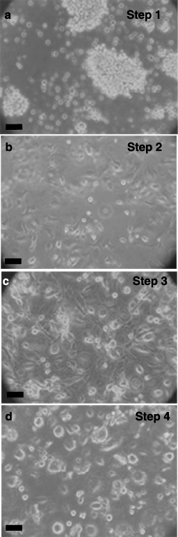
Differentiation of cord blood stem cells. Lineage‐negative cells were expanded for 1 week and then differentiated as described in ‘Methods’ and cell morphology shown with phase contrast microscopy. Step 1 (4 days, Panel a), aggregation in differentiation medium. Step 2 (6 days, Panel b), production of nestin‐positive, attached cells in serum‐free medium. Step 3 (6 days, Panel c), proliferation towards pancreatic lineage with basic fibroblast growth factor (bFGF). Step 4 (6 days, Panel d), differentiation to insulin‐ and C‐peptide‐containing cells by nicotinamide. Phase contrast micrographs are representative of at least five experiments. Scale bars, 25 µm.