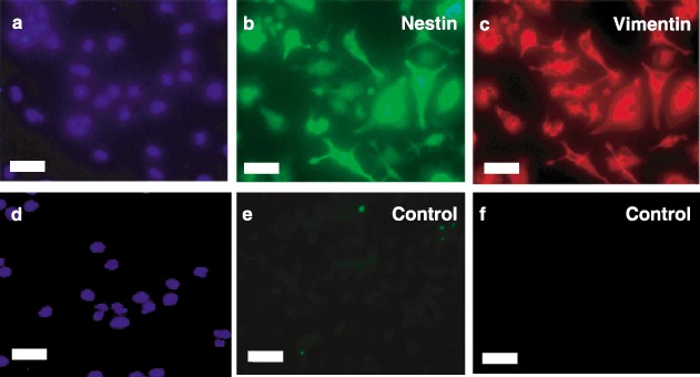
Figure 4.

Immunocytochemical characterization of nestin and vimentin expression. Lineage‐negative stem cells after serum‐free induced attachment in Step 2 were fixed and stained with antihuman nestin IgG (Panel b), antihuman vimentin immunoglobulin G (IgG) (Panel c) or control IgG (Panels e and f). Cells were then exposed to fluorescein isothiocyanate (FITC) (Panels b and e) – or Texas Red (Panels c and f) – conjugated secondary antibodies and nuclei stained with 4′,6‐diamidino‐2‐phenylindole (DAPI) (Panels a and d). Fluorescent micrographs are representative of at least three experiments. Scale bars, 25 µm.
