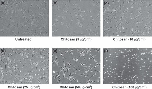
Figure 2.

hMSCs culture on tissue culture plates coated with different concentrations of chitosan. (a) Cells on untreated plates. (b–f) Cells on chitosan coated plates. (b–d) hMSCs attached and showed fibroblast like morphology on chitosan plates of coating density 5, 10 and 25 μg/sq. cm. There was uniform cell distribution on these plates. (e) Chitosan plate with a coating density of 50 μg/sq. cm also showed adherent hMSCs but there was non‐uniform cell distribution. (f) Chitosan plate with a coating density of 100 μg/sq. cm did not show any adherent hMSCs but there was formation of non‐adherent cell aggregates on this plate. A coating density of 25 μg/sq. cm which supported hMSCs adhesion was selected for further osteoblast differentiation experiments. Phase contrast micrographs with a total magnification of 40×.
