
Figure 1.
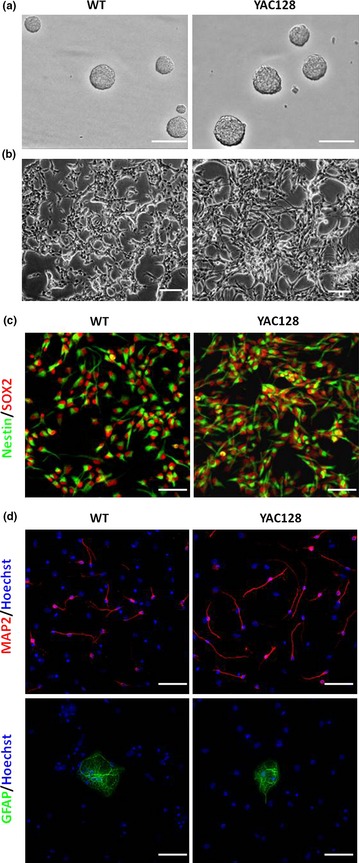
ANPC cultures and monolayer ANPC differentiation in vitro . (a) ANPCs were grown as neurospheres in a non‐coated six‐well plastic plate. Scale bar: 250 μm. (b) ANPCs were grown as monolayers in poly‐ornithine‐coated six‐well plastic plates. Scale bar: 250 μm. (c) Monolayer ANPCs were stained with Nestin (green) and SOX2 (red). Scale bar: 50 μm. (d) Monolayer ANPCs were able to differentiate into neurons (MAP2, red) and astrocytes (GFAP, green). Scale bar: 50 μm.