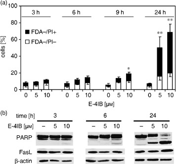
Figure 1.

E‐4IB induces apoptosis. (a) Thymidine‐synchronized HL60 cells were exposed to 5 or 10 µm E‐4IB for 3, 6, 9 and 24 h and FDA−/PI− apoptotic cells and apoptotic/necrotic FDA−/PI+ cell populations were calculated from flow cytometry measurements. Means of at least three independent experiments ± SD are shown. Statistical significance from the controls was evaluated, *P < 0.05, **P < 0.01. (b) Western blot analysis of PARP and FasL (50 µg protein lysate) is demonstrated. β‐actin was used as a loading control.
