
Figure 2.
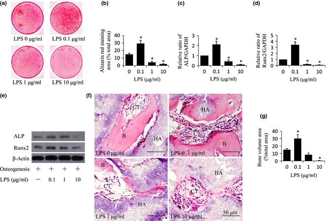
Porphyromonas gingivalis lipopolysaccharides ( Pg‐ LPS) at 0.1 μg/ml promoted and Pg‐ LPS at 10 μg/ml inhibited osteogenic differentiation of bone marrow mesenchymal stem cells ( BMMSC s). (a) Alizarin red staining indicating extracellular calcium deposition of BMMSCs after treatment with osteogenic media plus different concentrations (0, 0.1, 1 and 10 μg/ml) of Pg‐LPS, for 2 weeks. (b) Quantitative analysis of amounts of alizarin staining area as described in (a). (c, d) Relative mRNA levels of ALP (c) and Runx2 (d) in BMMSCs were measured by qRT‐PCR after treatment with osteogenic media plus different concentrations (0, 0.1, 1 and 10 μg/ml) of Pg‐LPS, for 7 days. (e) Western blot analysis of groups indicated in (a). (f) H&E staining of tissue samples from nude mice 8 weeks after subcutaneous implantation with BMMSCs pre‐treated with different concentrations (0, 0.1, 1 and 10 μg/ml) of Pg‐LPS for 12 h. Formation of bone (B) and connective tissue (CT) around HA‐TCP (HA) were indicated. Scale bar = 50 μm. (g) Quantitative analysis of amount of bone formation as described in (f). Results are representative of three independent experiments and are expressed as mean ± SD, n = 3 in (a–d), n = 5 in (e); statistical significance is shown as (*) P < 0.05, compared to BMMSCs without Pg‐LPS treatment. LPS, lipopolysaccharides from Porphyromonas gingivalis.