
Figure 2.
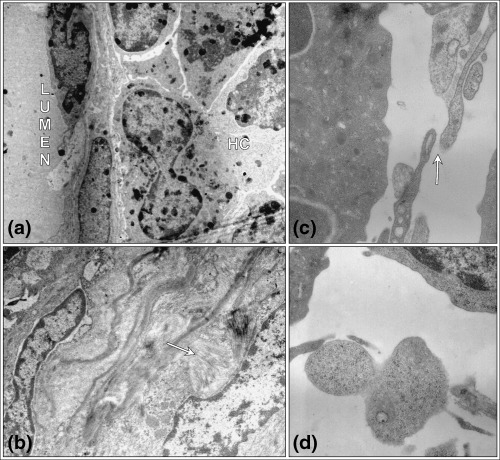
Ultrastructure of control mouse bone marrow on the days 2 and 4 of mobilization with G‐CSF plus CY. Endothelial cell resting on its discontinuous basement membrane, and cells of the haematopoietic compartment (HC) in the bone marrow (a). The destructive effect of cyclophosphamide on the 2nd day of mobilization – deposits of collagen fibres (arrow) are observed (b). Distance between endothelial cells (arrow) (c) and the passage of cells across the sinusoid fenestration (d) 4th day of mobilization. TEM (a) magnification × 4000; (b) magnification × 7500; (c) magnification × 20 000; (d) magnification × 15 000 (magnifications at time of photography).